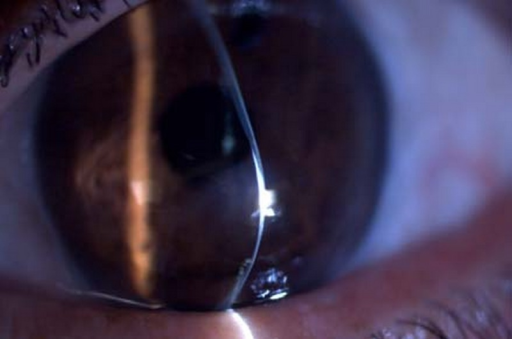

According to epidemiological studies about the incidence of PMD, this condition is considered as a rare condition, less common than other ectatic diseases, such as keratoconus, but more common than others, such as keratoglobus or posterior keratoconus. Furthermore, it may be considerably underestimated because PMD is often misdiagnosed as keratoconus due to its close resemblance in the clinical presentation. The incidence or prevalence of PMD is not clearly reported in bio-statistical studies. The term “pellucid” means clear and was used by the first time by Schlaeppi to denote the clarity of the cornea and the absence of any scarring, lipid deposition or vascularization, despite the presence of ectasia. However, there are several reports showing PMD cases with involvement of superior, and even temporal and nasal regions of the cornea. This condition most commonly involves the inferior cornea, with a thinning extending from the 4-o´clock to the 8-o´clock positions. Pellucid marginal degeneration (PMD) is a non-inflammatory and progressive ectatic corneal disease characterized by a narrow band of corneal thinning separated from the limbus by a relatively uninvolved area 1–2 mm in width. In addition, biomechanical and densitometric properties have been studied as complementary techniques to help in the diagnosis of PMD. New Scheimpflug imaging-based devices have shown the importance and usefulness of the pachymetric map for an appropriate diagnosis of PMD. Corneal topographic indices and the classical crab-claw topographic pattern cannot be used as the main tool to distinguish between PMD and keratoconus.

Slit-lamp examination is very useful to distinguish PMD from other corneal ectatic disorders with inflammatory nature.
PMD usually starts later in life than keratoconus and progresses slower than keratoconus. It is a rare corneal disorder that shares many clinical characteristics with other corneal ectasias, such as keratoconus, keratoglobus or Terrien marginal degeneration. Pellucid marginal degeneration (PMD) is a non-inflammatory ectatic corneal disease characterized by a narrow band of corneal thinning separated from the limbus by a relatively uninvolved area 1–2 mm in width.


 0 kommentar(er)
